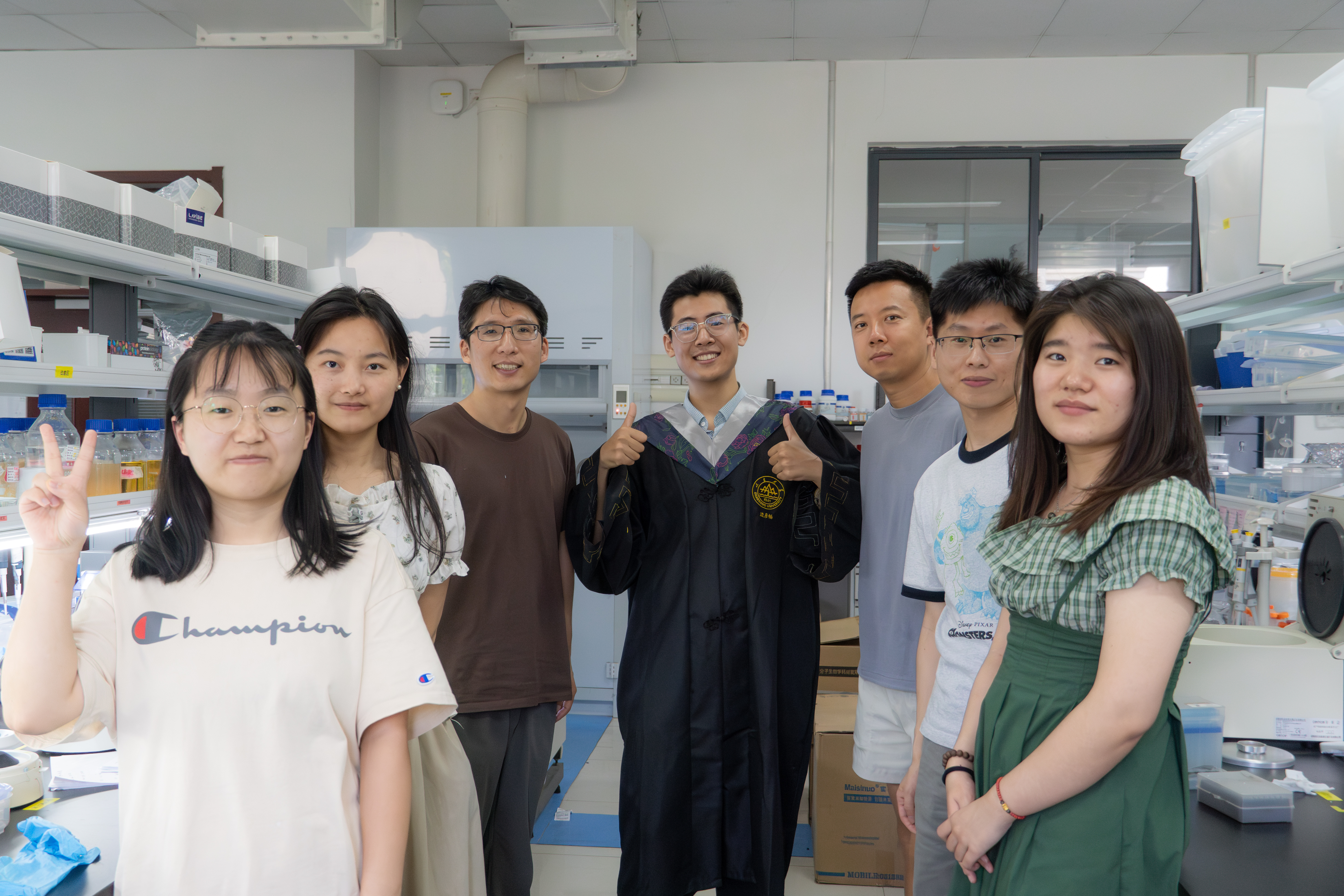
Yanchang Bian

Yanchang Bian

I am a Junior Specialist in the laboratory of Prof. Dengke Ma at the University of California, San Francisco. I am interested in how cells organize molecular components across space and time, and how their interactions give rise to coordinated, adaptable behavior.

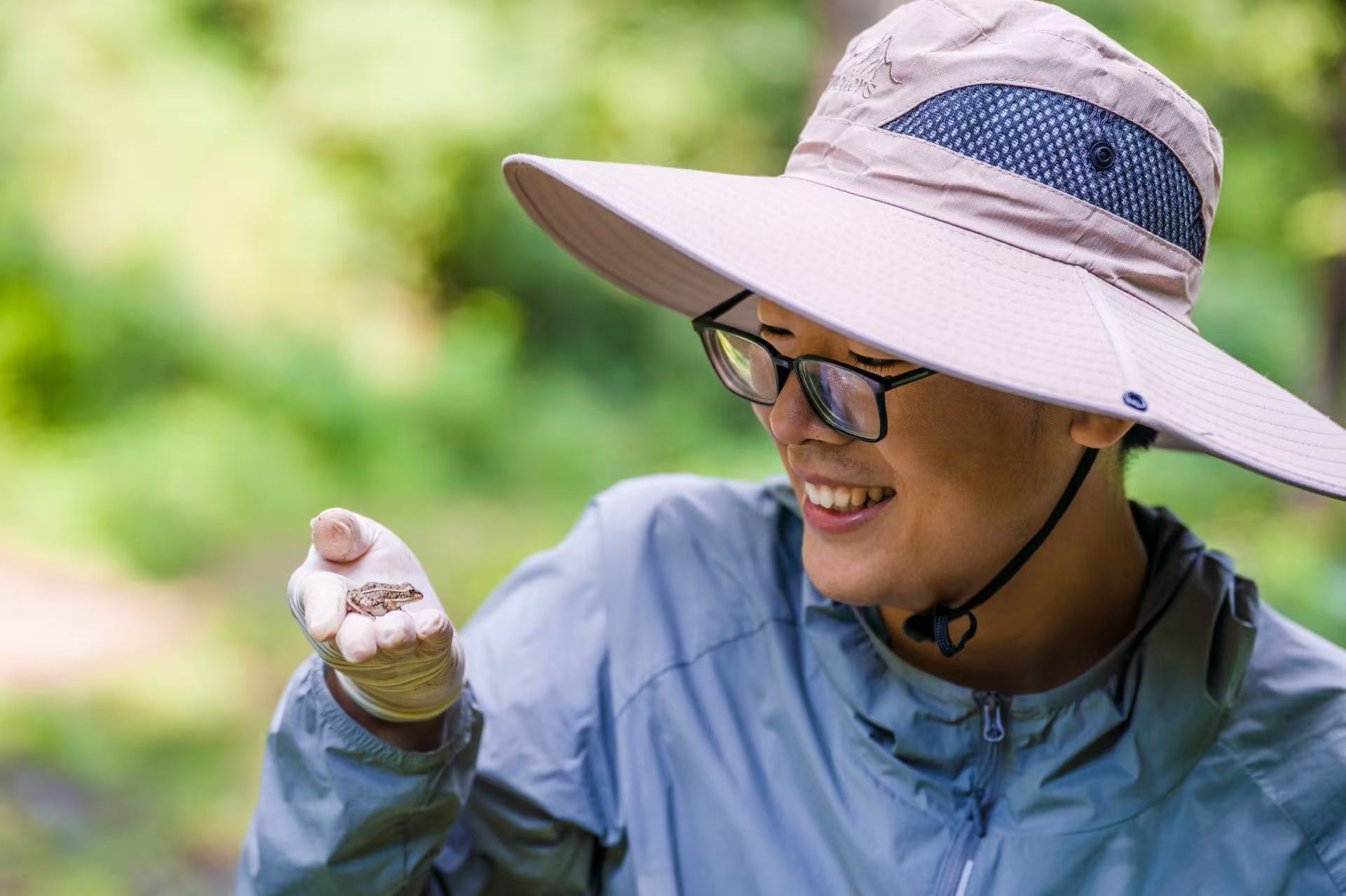
Yanchang Bian observing a small frog resting on his gloved hand outdoors

Publications & manuscripts

Molecular CellManuscript under review
First round
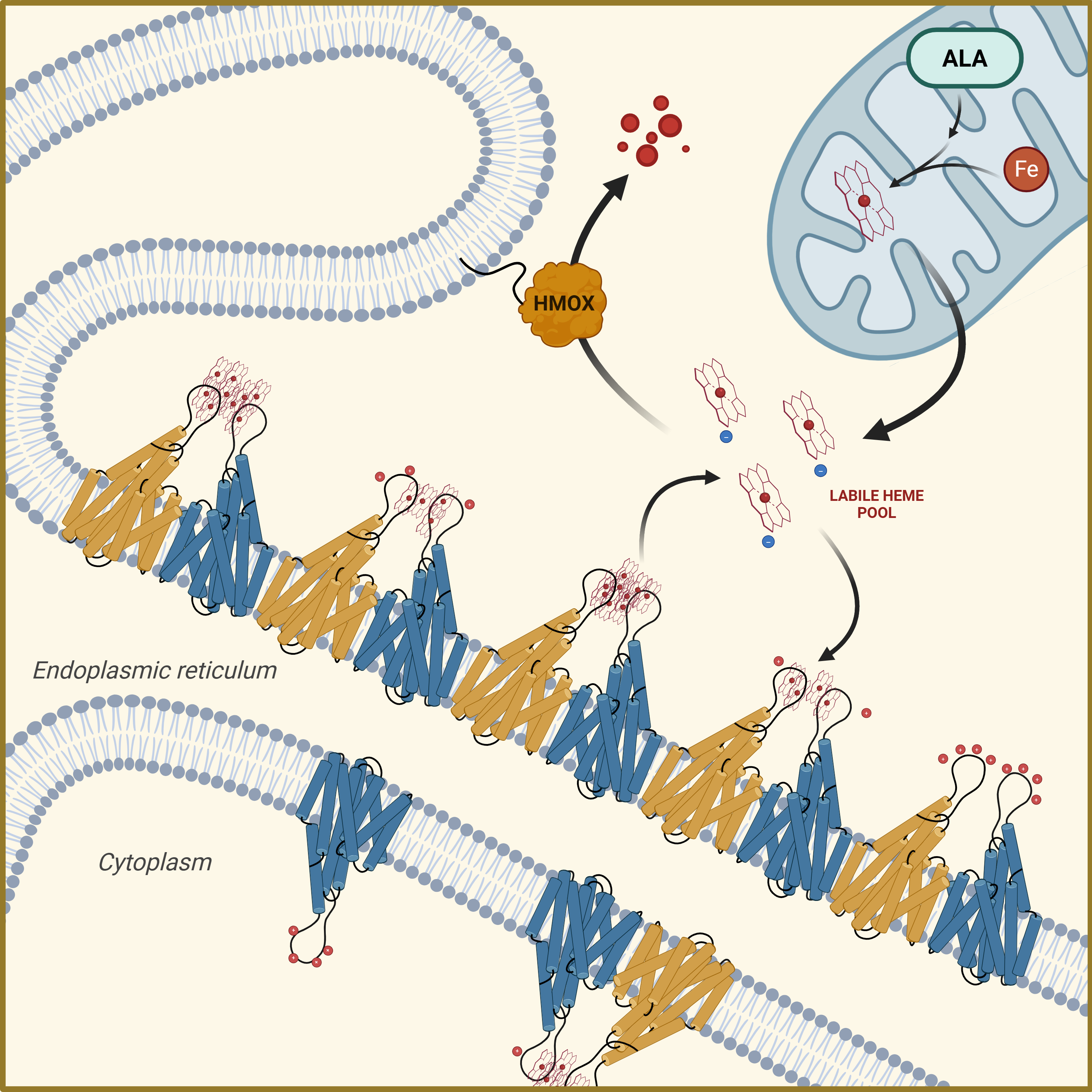
Proposed TMEM161B heme-buffering model at the endoplasmic reticulum

Linear assembly of TMEM161B reveals a conserved mechanism for intracellular heme management

Yao, J., Bian, Y., Xu, S., Liang, S., Ji, Z., Liu, J., Chen, Y., Rao, Z., Ma, D., and Lou, Z.

Research poster (PDF)
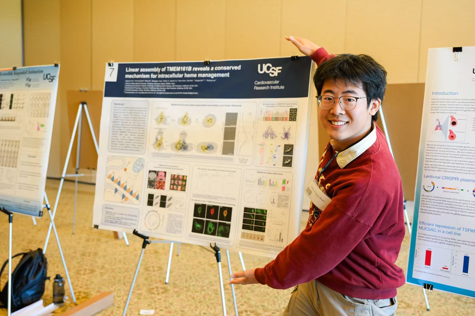
20260925 UCSF BMS Retreat
Presenting the TMEM161B poster at the UCSF BMS Retreat on September 25, 2026
Abstract

Heme is essential for numerous cellular processes and is dynamically trafficked within cells, yet its labile form is a potent pro-oxidant and signaling molecule whose concentration must be held within a narrow physiological range. How cells achieve this control remains poorly understood. Here we identify the conserved endoplasmic reticulum (ER) membrane protein TMEM161B as a key regulator of the labile heme pool. We report cryo-EM structures of human TMEM161B in monomeric and dimeric states that further assemble into extended filamentous arrays through two distinct oligomerization interfaces. The cytosolic loop of each dimer forms a pseudo-C2-symmetric loop clamp that coordinates up to 8–9 heme molecules, representing a previously undescribed mode of membrane-associated heme binding. Using a genetically encoded cytosolic heme sensor, we show that TMEM161B overexpression reduces the labile heme pool whereas knockdown elevates it, accompanied by corresponding heme-responsive transcriptional changes. In C. elegans, loss of the TMEM161 ortholog strongly suppresses the hrg-1p::GFP reporter, consistent with an elevated available heme pool, demonstrating that this buffering function is conserved from nematodes to humans. Together, these findings define TMEM161B as a membrane-embedded heme buffer that assembles into linear arrays on the ER, and raise the possibility that the labile heme pool is spatially compartmentalized across organelle membranes to a greater extent than currently recognized.
Cell Reports23 Jun 2026
Vol 45, Issue 6
Article 117489

A conserved antioxidant defense at the endoplasmic reticulum membrane

Ji, Z., de Belly, H., Pandey, T., Wang, B., Tang, Y., Yao, J., Xu, S., Li, K., Bian, Y., Guang, S., et al.

Abstract

Oxidative protein folding in the endoplasmic reticulum (ER) is essential for eukaryotic cells yet generates hydrogen peroxide (H2O2), a reactive oxygen species. The ER-transmembrane protein that supports ER proteostasis and guards the cytosol for antioxidant defense remains unidentified. Here, we combine AlphaFold2 and functional screens in C. elegans to discover a previously uncharacterized and evolutionarily conserved protein ERGU-1 that fulfills these roles. Deleting ERGU-1 upregulates H2O2 and NRF2/SKN-1-dependent gene expression. ERGU-1 deficiency also impairs organismal reproduction and behavioral responses to H2O2. Both C. elegans ERGU-1 and human homolog TMEM161B localize to ER membranes, forming reticular networks. Human and Drosophila homologs of ERGU-1 rescue C. elegans mutant phenotypes, demonstrating ancient and conserved functions. In addition, purified ERGU-1 and TMEM161B exhibit redox-modulated oligomeric states. Together, our results reveal an ER-membrane-specific machinery, suggesting a conserved mechanism for maintaining ER redox homeostasis and proteostasis in animal cells.

Published abstract · Source
Science Advances24 Jul 2026
Vol 12, Issue 30
Article eaef3219

A genetically encoded sensor of ionic stress links cellular proton dynamics to sleep

Ji, Z., Wang, B., Ma, Z., Verbakel, L., Liu, J., Bian, Y., Zeng, W., Chung, C.-I., Wei, S., et al.

Abstract

Although biosensors for specific cellular ions are widely available, real-time monitoring of overall ionic strength in living organisms remains challenging. Here, we present a genetically encoded nuclear translocation ionic sensor (GENTIS) that enables direct visualization of ionic stress in vivo. Using this sensor alongside longitudinal tracking via an automated microfluidic platform, we find that Caenorhabditis elegans larvae experience highly synchronized, rhythmic elevations in intestinal ionic strength during the molt, a stage during which developmentally timed sleep occurs. Cytosolic proton accumulation through inhibition of vacuolar-type adenosine triphosphatases (V-ATPases) triggers GENTIS nuclear translocation and evokes behavioral quiescence, characterized by reduced feeding, locomotion, and activation of sleep-active neurons. Apical membrane V-ATPases naturally undergo disassembly during molting and stress, conditions that cause proton accumulation and sleep. Notably, this proton-linked sleep is suppressed by proton buffering with ammonium. Together, these findings establish GENTIS as a powerful tool for tracking ionic strength dynamics in vivo and reveal that proton ionic rhythms contribute to the regulation of sleep.

Published abstract · Source

Background

Research experience

University of California, San Francisco

Junior Specialist · Prof. Dengke Ma
Cardiovascular Research Institute

Project details

I study membrane and metabolic remodeling during hypometabolic states in C. elegans. During liquid-induced suspended animation, I observed loss of V-ATPase signal from the intestinal apical membrane. Blue native PAGE revealed fewer fully assembled complexes and increased free V₁ sector, supporting V₁–V₀ disassembly. Similar loss of apical signal occurs during lethargus.

I also investigate a candidate regulator of fatty-acid oxidation identified through proteomics. After purifying the candidate protein, ETFA/B, and medium-chain acyl-CoA dehydrogenase, I established a DCIP-based electron-transfer assay and observed concentration-dependent inhibition by the candidate. Ongoing work tests its relevance in vivo. In parallel, I continue studying TMEM161B-dependent heme homeostasis using dietary heme supplementation and C. elegans genetics.

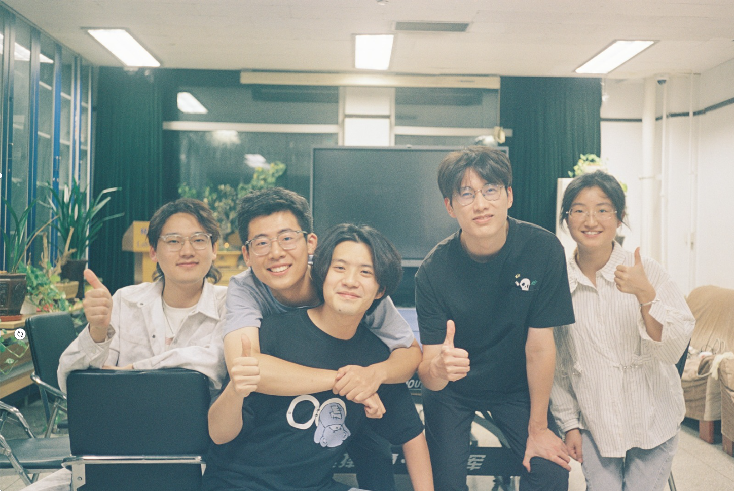
With the Ma lab at UCSF

Tsinghua University

Research Assistant · Prof. Zhiyong Lou
School of Basic Medical Sciences

Project details

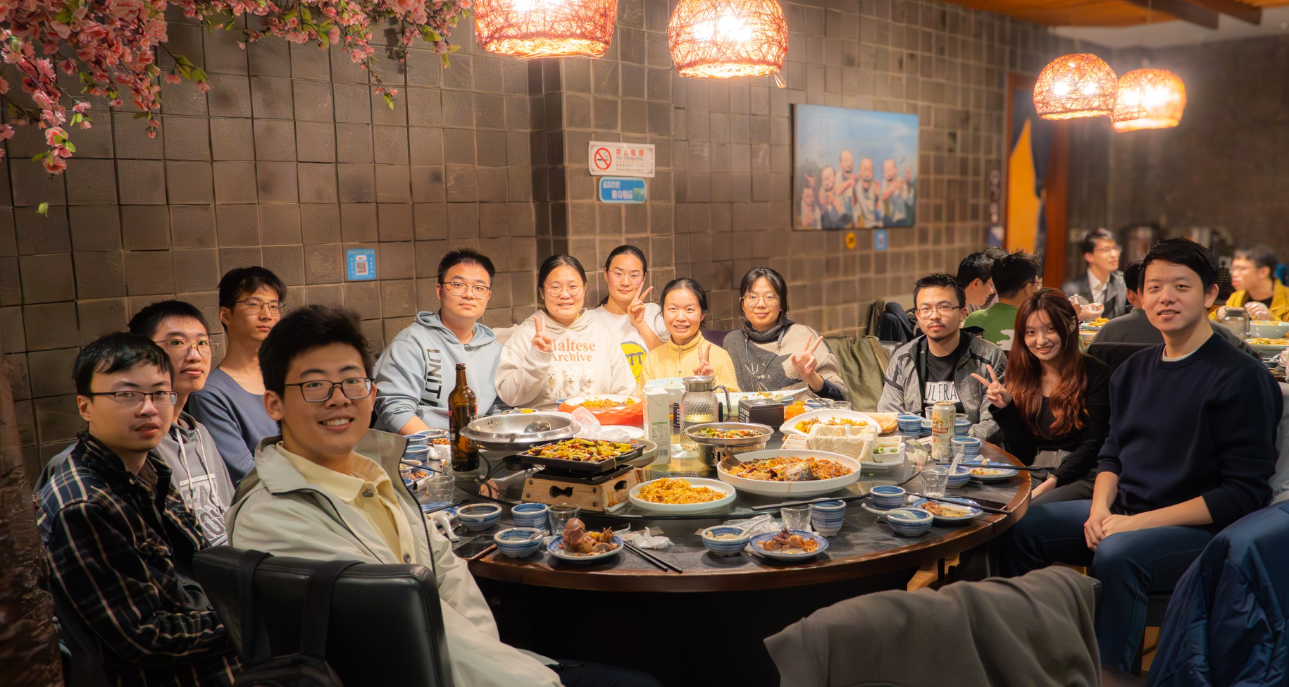
I used cryo-EM to investigate the structure and higher-order assembly of TMEM161B. The protein forms C2-symmetric homodimers that further assemble into linear oligomers. Additional density near a cytosolic loop led us to investigate heme binding.

Mutational analysis supported the loop’s role in heme binding, while quantitative ITC measurements established a stoichiometry of approximately four to five heme molecules per TMEM161B monomer. This work connected structural assembly with a potential mechanism for cellular heme management.

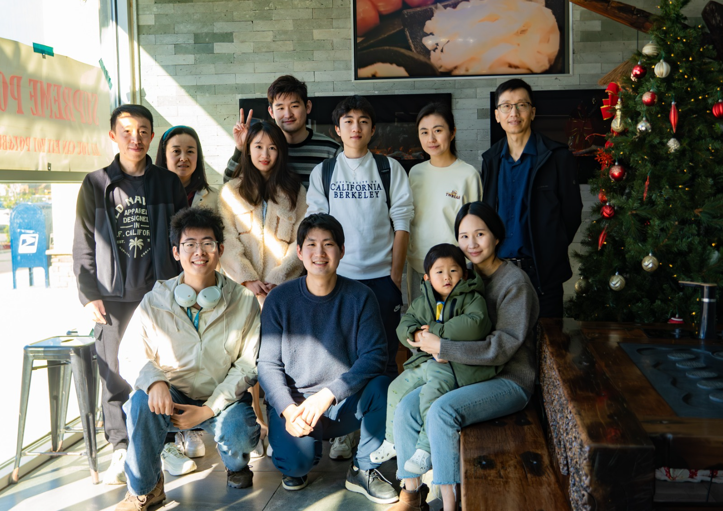
With the Lou lab at Tsinghua University

Tsinghua University

Undergraduate Researcher · Prof. Yinqing Li
IDG/McGovern Institute for Brain Research

Project details

I developed an Escherichia coli split-adenylate-cyclase dual-hybrid system to quantify interactions among transcription-factor intrinsically disordered regions, using flow cytometry and amplicon sequencing.

I validated the system with FUS, directly observed FUS condensates in E. coli, and generated a 16 × 16 IDR interaction matrix that recovered established pairs, including MED1–BRD4.

With the Li lab at Tsinghua University

Shandong University

Undergraduate Researcher · Prof. Guang Zhao
State Key Laboratory of Microbial Technology

Project details

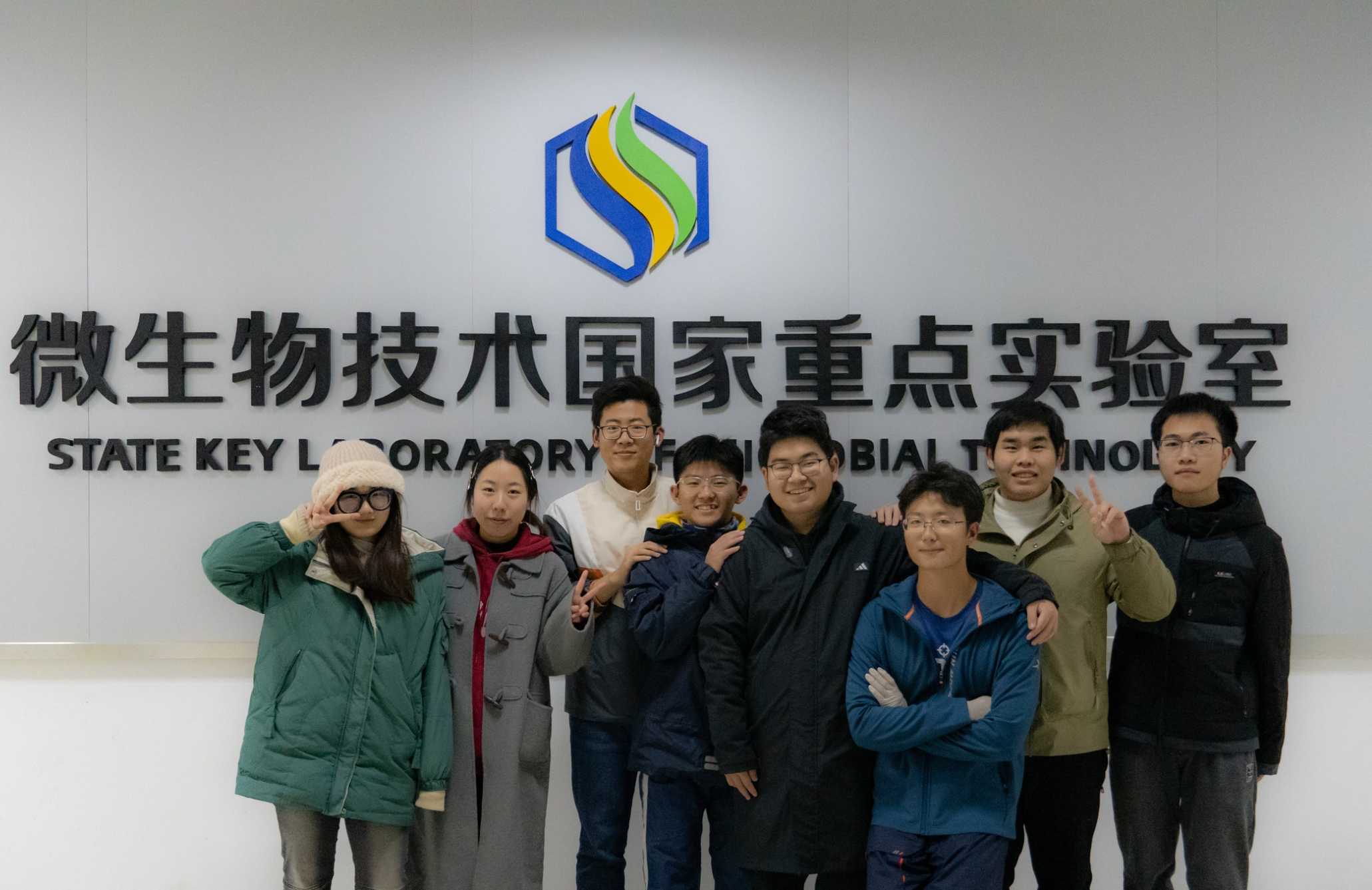
I characterized acetylation of the transcription factor ArcA by Pat and acetyl phosphate, and its deacetylation by CobB. Using site-directed mutagenesis, electrophoretic mobility shift assays, and qRT-PCR, I investigated how acetylation affected its regulatory activity.

Acetylation of specific lysine residues reduced ArcA DNA-binding activity and altered target-gene expression, linking a biochemical modification to transcriptional regulation.

With the Zhao lab at Shandong University
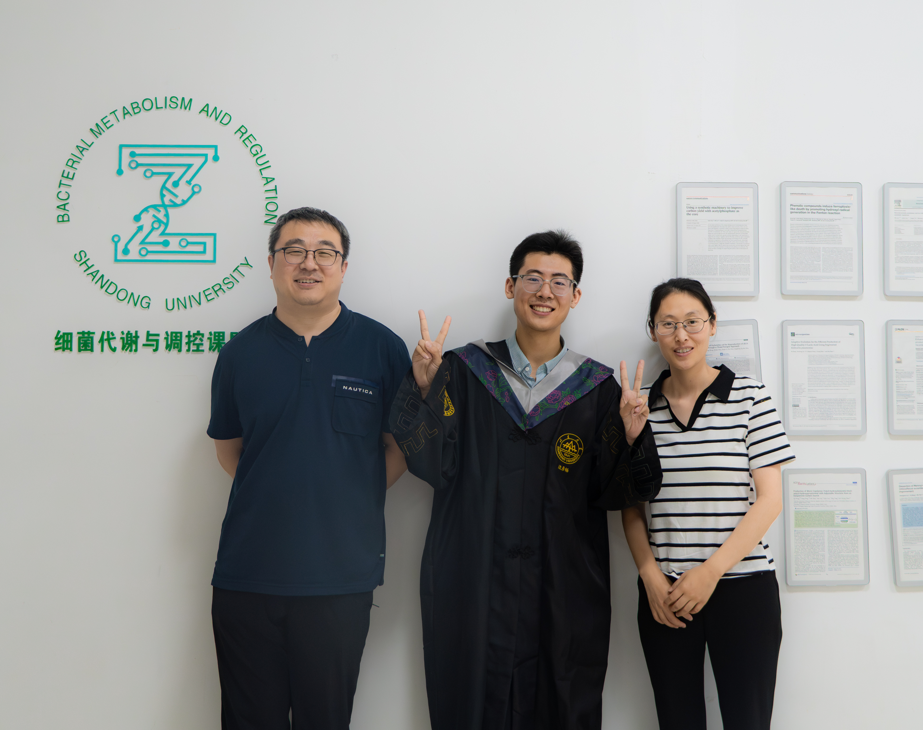
With Prof. Guang Zhao and Prof. Min Liu

Westlake University

Undergraduate Researcher · Prof. Zhubing Shi
School of Life Sciences

Project details

I purified PAF1 and P-TEFb transcription complexes for biochemical reconstitution, using Ni-NTA and Strep-tag affinity purification, size-exclusion chromatography, and anion-exchange chromatography.

This project provided training in ÄKTA chromatography and cryo-electron microscopy workflows.

Shandong University

Undergraduate Researcher · Prof. Ye Hong
School of Life Sciences

Project details

I investigated NATH-10 function in C. elegans reproduction and meiosis using RNA interference, auxin-inducible degradation, yeast two-hybrid assays, and immunofluorescence.

NATH-10 localized predominantly to the nucleolus, and we detected no interaction with the proposed chromosome-axis proteins. The project received the National First Prize in the China Undergraduate Life Sciences Contest.

With the Hong lab at Shandong University

Research interests

I am most interested in how cells organize dynamic molecular components across space and time, and how local interactions give rise to stable yet adaptable cellular behaviors. I am particularly drawn to molecular localization, intracellular transport, higher-order protein assembly, and organelle and cytoskeletal organization. Rather than asking only what a protein does, I want to understand where it acts, how it gets there, and how microscopic rules scale into cell-level function.

I still vividly remember the first time I watched living cells through a 60× objective: mitochondria twisting through the cytoplasm like worms, while lysosomes moved rapidly through dividing cells. Life is alive. Cells are alive. What fascinated me most was that all this motion did not produce chaos. Molecules and organelles constantly move, reshape, and redistribute, yet the cell remains organized enough to function. I want to understand the rules that make this possible. My work on TMEM161B brought me to the same question at a molecular scale: its function appeared to depend not only on what it could bind, but also on how it assembled and was organized on the ER membrane.

For my PhD, I hope to combine mechanistic biochemistry and cell biology with quantitative approaches such as advanced imaging, single-molecule methods, Cryo-EM, and mathematical modeling. I hope to join a PhD program that provides interdisciplinary training in experimental and quantitative approaches, helping me connect molecular mechanisms with cell-level behavior.

In the long term, I hope to contribute to a theoretical understanding of how complex cellular behaviors emerge from molecular interactions. Even if powerful AI models can reproduce or predict cellular behavior, I do not think prediction alone amounts to understanding. Within my lifetime, I hope to see biology uncover interpretable principles that explain why such behaviors emerge, and I want my research to help bridge molecular mechanisms with those broader principles.

Techniques

Protein biochemistry
Recombinant expression and purification, affinity and size-exclusion chromatography, ion-exchange chromatography, ITC, blue native PAGE, EMSA, co-immunoprecipitation, and enzyme assays.
Molecular biology
Molecular cloning, site-directed mutagenesis, qRT-PCR, CRISPR–Cas9, RNA interference, amplicon-library preparation, and lentiviral packaging and transduction.
Cells & imaging
Mammalian cell culture, flow cytometry, immunofluorescence, live-cell and spinning-disk confocal microscopy. T12 negative-stain electron microscopy.
Analysis
Python, RNA-seq analysis, Scanpy, FlowJo, ImageJ/Fiji, GraphPad Prism, and SnapGene.

Outside the lab

I enjoy biological observation, nature photography, hiking, and documenting biodiversity.

My observations on iNaturalist

CONTACT

+1 (628) 726-4851

San Francisco, California

My field observations

—Observations
—Species

Loading observations from iNaturalist…

Explore all observations on iNaturalist
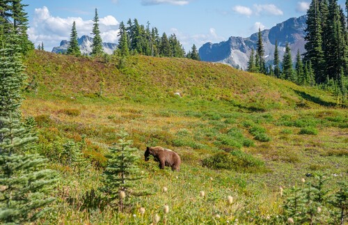
American Black Bear
American Black Bear
(c) ingester, some rights reserved (CC BY-NC)
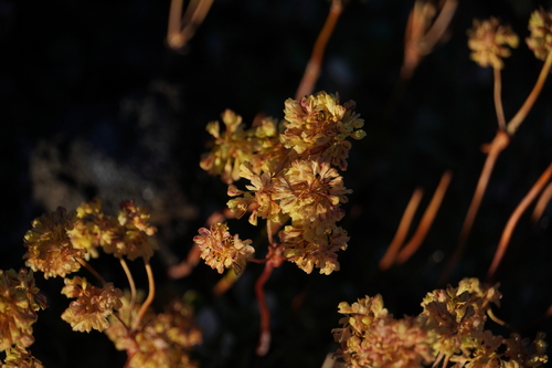
marumleaf buckwheat
marumleaf buckwheat
(c) ingester, some rights reserved (CC BY-NC)
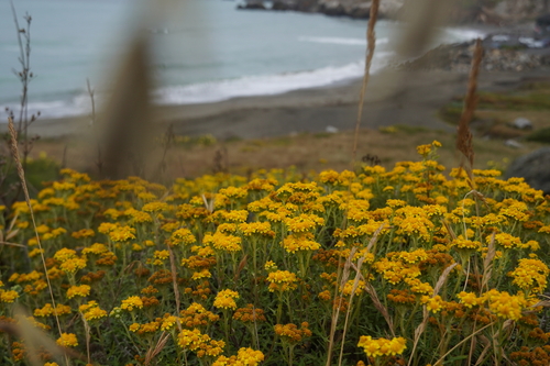
seaside woolly sunflower
(c) ingester, some rights reserved (CC BY-NC)
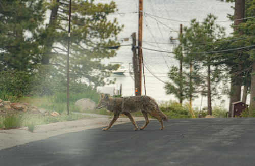
Coyote
Coyote
(c) ingester, some rights reserved (CC BY-NC)
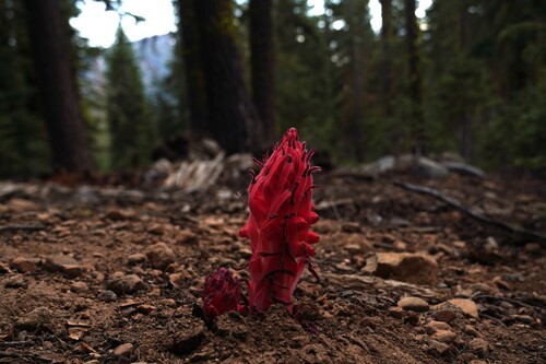
Snowplant
Snowplant
(c) ingester, some rights reserved (CC BY-NC)
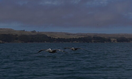
Brown Pelican
Brown Pelican
(c) ingester, some rights reserved (CC BY-NC)